Patient-specific ‘avatars’ for paediatric brain cancer are becoming more realistic — but they remain mostly research tools
Patient-specific ‘avatars’ for paediatric brain cancer are becoming more realistic — but they remain mostly research tools
In paediatric oncology, few fields make the shortcomings of current medicine as visible as brain cancer. Even with advances in molecular diagnosis, neurosurgery, radiotherapy and targeted treatment strategies, many paediatric brain tumours remain devastatingly hard to treat. Part of the difficulty lies in the tumours themselves: they are biologically diverse, often highly invasive and deeply shaped by the unusual environment of the developing brain.
That is why the idea of creating an “avatar” of a child’s tumour has become so compelling. In practice, an avatar model is a patient-specific system built from the tumour’s own cells and designed to reproduce, as faithfully as possible, how that cancer behaves in the body. The more realistic the model, the more useful it may become for understanding tumour biology, testing drugs and, eventually, supporting more precise treatment decisions.
The evidence provided here supports the idea that this once futuristic concept is becoming increasingly feasible in paediatric brain cancer. In particular, organoids and orthotopic patient-derived xenografts are emerging as more realistic tools for studying how these tumours grow, infiltrate and respond to therapy. But the crucial caveat is that these systems remain, for now, largely preclinical research platforms rather than standard tools used routinely in paediatric cancer care.
Why simple models are no longer enough
For years, much of cancer research relied on relatively simple laboratory systems such as tumour cell lines grown in flat culture dishes. Those models were useful, but they came with clear limitations. Over time, cells adapted to artificial lab conditions can lose some of the features that made the original tumour clinically important.
That problem is especially serious in brain cancer. Tumours do not exist in isolation. They interact with surrounding brain tissue, immune cells, blood vessels and structural constraints that shape how they grow and spread. A model that strips away too much of that complexity may be easier to study, but it can also become less clinically relevant.
This is why more advanced three-dimensional systems have attracted so much interest. A broader review of 3D cell culture approaches included in the supplied literature concludes that organoids and related patient-specific models better preserve histologic, transcriptional, mutational and heterogeneous features of brain tumours than simpler systems do. That matters because fidelity is the entire point of an avatar. If a model does not resemble the real tumour closely enough, its usefulness for precision oncology is limited.
Organoids are beginning to capture tumour behaviour, not just tumour cells
One of the most striking developments in the supplied evidence is a recent human-cell-based fusion organoid model for diffuse midline glioma, one of the most aggressive and difficult paediatric brain tumours.
According to the material provided, this model recreated diffuse tumour infiltration and interactions with brain immune cells. That is a major step forward. Diffuse midline glioma is so lethal in part because it does not behave like a neatly contained mass. It infiltrates delicate brain structures, making surgical removal impossible and complicating treatment in ways that simpler lab models often fail to capture.
An organoid that can reproduce at least part of that infiltrative behaviour starts to look much more like a meaningful tumour avatar rather than a basic cluster of cancer cells. It becomes a system for studying not just what the tumour is, but what it does.
That distinction matters in precision oncology. Understanding tumour behaviour — how it invades, how it interacts with the local immune environment and how it responds to therapeutic pressure — is often more clinically useful than cataloguing mutations alone. If organoid systems can model those dynamics with increasing realism, they could become powerful tools for both mechanistic discovery and treatment testing.
Orthotopic patient-derived xenografts remain a major part of the picture
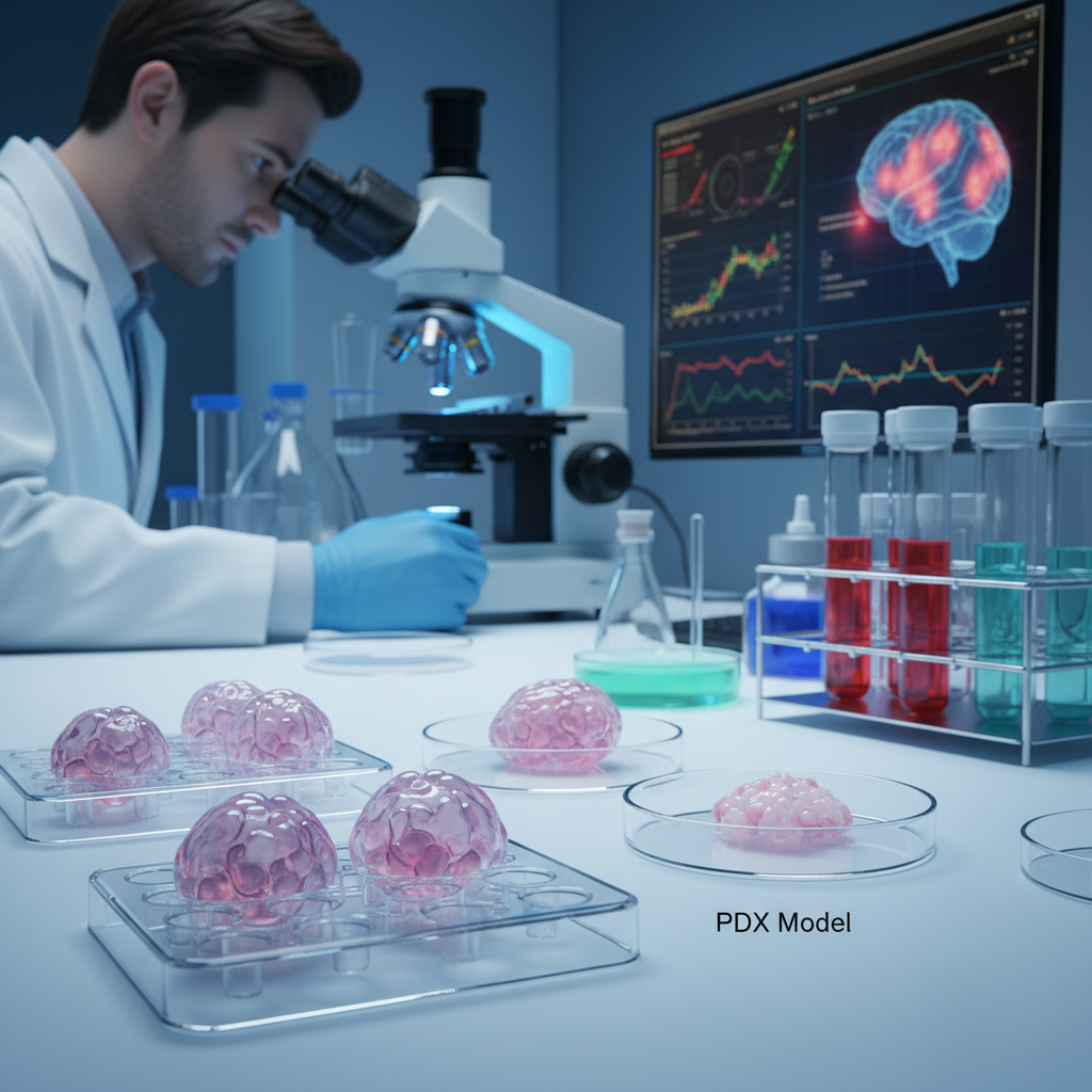
Organoids are not the only platform driving this shift. Orthotopic patient-derived xenograft models, or PDX models, are already an established part of preclinical paediatric brain tumour research.
In these systems, tumour cells taken from a patient are implanted into an anatomically relevant site, typically the brain of a laboratory animal, so the cancer can grow in an environment that is more biologically meaningful than a dish. In paediatric intracranial tumours, this helps researchers reproduce tumour growth and behaviour under conditions that better reflect the real disease.
That relevance matters. Brain tumours are shaped by location, tissue architecture, local signalling, drug penetration and the challenges of the brain environment itself. Orthotopic PDX models are useful precisely because they preserve more of that context than simpler models can.
The supplied evidence notes that these models are already being used to support preclinical therapeutic testing. That makes them one of the strongest existing examples of how avatar-like approaches are already contributing to paediatric neuro-oncology research.
Why the ‘avatar’ idea now feels increasingly credible
Taken together, the supplied references support a fairly clear conclusion: clinically meaningful paediatric brain tumour avatars are becoming more plausible.
That does not mean researchers have created a perfect copy of a patient’s cancer. It means the field is moving closer to models that preserve enough of a tumour’s structure, molecular identity, heterogeneity and behaviour to make them genuinely useful. Different platforms contribute different strengths. Organoids may better capture certain cellular interactions and tissue-like organisation. Orthotopic xenografts may provide more realistic growth conditions inside a brain-relevant environment. Reviews of 3D systems suggest that both are substantial advances over older, simpler approaches.
This is especially important in paediatric brain cancer because these tumours are often rare, biologically distinct and difficult to study at scale. Better models do not just improve lab science in the abstract; they may help researchers identify which therapeutic ideas are worth pursuing in diseases where every patient sample is precious.
What this could mean for treatment in the future
The long-term appeal of avatar models is obvious. In theory, a child’s tumour cells could be used to build a personalised model, multiple therapies could be tested against that model, and clinicians could use the results to make more informed treatment choices.
That is the precision-oncology dream. But it is not yet the everyday clinical reality.
At present, the evidence is much stronger for avatar models as research and preclinical testing tools than as direct guides for patient care. They are helping scientists ask better questions, understand tumour mechanisms more clearly and test therapies in systems that better resemble real disease. That alone is important. In oncology, one reason promising drugs fail is that the models used to select them were not realistic enough. If better avatar systems improve that filtering process, they could have major indirect clinical value even before they become bedside tools.
The limits are still substantial
The first limitation is the type of evidence. Most of what is supplied here remains preclinical and focused on model development. That is not trivial — it is how translational science progresses — but it is not the same thing as proving direct clinical benefit for children currently undergoing treatment.
The second limitation is complexity. These systems are technically demanding, resource-intensive and not yet standardised across routine paediatric oncology practice. Building a high-quality organoid or patient-derived xenograft requires specialised infrastructure, expert teams and viable tumour material. That alone makes widespread clinical use difficult.
There are also biological limits. Even advanced organoids and xenograft models still cannot fully recreate the complete human tumour microenvironment, the systemic immune response, the full effects of radiotherapy and chemotherapy, or every feature of tumour evolution inside a patient. They are more realistic than older models, but they are still models.
Then there is the issue of time. For an avatar to directly shape treatment decisions, it has to be created, characterised and tested quickly enough to matter clinically. In aggressive paediatric brain tumours, that timeline can be very short. A model that is highly accurate but too slow to inform care may still be valuable for research, while offering less immediate benefit to the child from whom it was derived.
The most immediate impact may be on the quality of research
For now, the most realistic near-term value of these avatar systems may be that they improve the quality of preclinical science. That may sound modest, but it is not.
In a field where therapeutic progress can be painfully slow, using models that better reflect real tumours could help researchers discard weak strategies earlier and prioritise more promising ones. It could also improve understanding of why certain treatments fail, why some tumours invade so aggressively and how tumour cells interact with the surrounding brain.
For rare paediatric cancers, this matters even more. When patient numbers are limited, every high-quality model becomes more valuable. A better avatar can mean more biologically meaningful insight from a very small amount of precious tumour tissue.
A promising step, not a finished revolution
The phrase “avatar model” can sound futuristic, but in paediatric brain cancer it is becoming less of a metaphor and more of a realistic scientific goal. The supplied evidence supports that shift. Human-cell-based organoid systems are beginning to recreate key features such as diffuse infiltration and interactions with brain immune cells. Orthotopic patient-derived xenografts are already useful for modelling tumour growth in brain-relevant environments. Broader reviews of 3D systems reinforce that these approaches preserve more of what makes real tumours clinically important.
That is enough to justify cautious optimism. Paediatric brain tumour avatars are becoming increasingly realistic, and increasingly useful.
But the most honest conclusion is still a restrained one. These models are not yet a near-universal clinical reality, and they should not be presented as if personalised avatar-guided treatment is already standard care. Right now, they are primarily sophisticated research tools that may strengthen precision oncology by making tumour biology and treatment testing more realistic.
That may not be the final destination. But it is a meaningful step towards it.