AI may turn fat around the heart into a new cardiovascular risk clue — but for now it looks more like an add-on than a replacement
AI may turn fat around the heart into a new cardiovascular risk clue — but for now it looks more like an add-on than a replacement
Cardiovascular risk prediction has long relied on a familiar set of ingredients: age, blood pressure, cholesterol, diabetes, smoking, family history and, more recently, some imaging tools such as coronary artery calcium scoring. Now a newer line of research is trying to extract even more information from CT scans that many patients are already undergoing: the fat that sits around the heart and around the coronary arteries.
At first glance, that may sound like a technical detail. But the idea is gaining traction for a reason. Epicardial fat and perivascular fat do not appear to be biologically passive tissue. They may reflect inflammatory, metabolic and vascular processes that are relevant to atherosclerosis, remodelling and future cardiovascular events.
This is where artificial intelligence enters the picture. Rather than relying on slow, labour-intensive manual measurements, AI systems can quantify these fat depots automatically on imaging. In doing so, they may turn an underused anatomical feature into a useful biomarker. The supplied evidence supports that core idea reasonably well. What it does not support is the notion that this is about to displace standard cardiovascular risk tools.
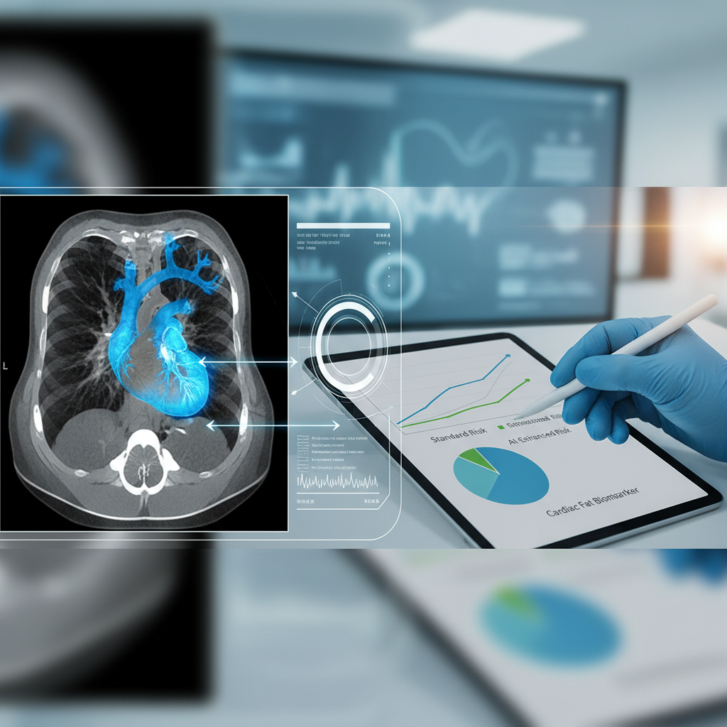
What exactly AI is measuring
When people hear about “heart fat”, it can sound vague. In practice, the literature supplied here focuses mainly on two related structures.
The first is epicardial adipose tissue, the fat located between the heart muscle and the pericardium, lying close to the coronary arteries. The second is perivascular fat, the tissue that surrounds blood vessels and may contain signals about the local inflammatory environment.
These tissues matter because they may not be inert. They can produce inflammatory mediators, interact with nearby vascular structures and reflect cardiometabolic states that conventional risk factors do not fully capture.
Until recently, the problem was practical. Measuring these fat compartments accurately and consistently at scale was not easy. AI changes that by making the process much more automated.
What the studies actually show
One of the strongest references in the supplied set describes a deep-learning approach for automated epicardial adipose tissue assessment. The system showed strong agreement with human measurement and found that epicardial fat independently predicted myocardial infarction, stroke, atrial fibrillation and all-cause mortality.
That matters for two reasons. First, it suggests that AI can extract a structurally meaningful measurement with enough fidelity to approach expert human assessment. Second, the measurement was not just technically feasible; it also carried prognostic value for clinically important outcomes.
Another study focused on a machine learning-derived radiomic profile of perivascular fat. In that work, the perivascular fat signal significantly improved prediction of major adverse cardiac events beyond traditional risk factors, coronary calcium score, stenosis severity and high-risk plaque features seen on coronary CT angiography.
Together, those findings support the idea that cardiac and perivascular fat contain useful risk information that current imaging workflows may not fully exploit — and that AI can help surface it.
The promise is refinement, not reinvention
The most balanced way to interpret these findings is not as a replacement for existing cardiovascular risk models, but as a possible refinement of them.
Preventive cardiology often operates in grey zones. Many patients fall into intermediate-risk categories where the practical question is not whether they have some risk, but whether they carry enough risk to justify more intensive prevention, closer follow-up or additional testing.
That is where imaging biomarkers may prove most valuable. If AI-derived measurements of epicardial or perivascular fat improve discrimination, even modestly, they may help separate out which patients within apparently similar groups actually face higher risk.
That distinction is important. Improving predictive accuracy is not the same as creating a universal new best test. Sometimes it means adding a clinically useful layer of nuance in selected settings.
Why fat around the heart may matter biologically
Part of the appeal of this research lies in the biology. Fat around the heart and coronary vessels may do more than simply reflect body composition. It may be part of a local environment tied to inflammation, vascular remodelling and atherosclerotic activity.
That is one reason perivascular fat, in particular, has attracted so much interest. Instead of functioning only as a marker of general adiposity, it may offer a more localised readout of what is happening in and around the vessel wall.
If that interpretation continues to hold up, AI would not merely be measuring fat. It would be extracting an indirect signal of vascular biology.
Where the caution begins
Even so, the evidence provided comes with important limits. The most recent review in the supplied set notes that perivascular fat measures may add only modest predictive discrimination in large datasets.
That word — modest — matters a great deal. In biomarker research, statistically significant improvements do not automatically translate into clinically transformative ones. A model can become somewhat better at forecasting events without materially changing treatment decisions or outcomes.
There is also a setting issue. Most of the evidence comes from CT-based imaging studies rather than broad population-wide screening settings. That means the findings are promising, but they are not yet the same as showing usefulness across routine care at scale.
Better prediction is not automatically better care
This is one of the most important distinctions in the story.
There are really three different levels of evidence in play. First, a biomarker may correlate with risk. Second, it may improve a prediction model. Third, using that biomarker in practice may improve what happens to patients.
The supplied references speak fairly well to the first two levels. They do not yet establish the third. Improved predictive performance does not automatically mean clinicians will make better decisions, or that patients will have fewer heart attacks, fewer strokes or lower mortality because the marker was measured.
So any claim that this is already changing patient outcomes would be premature.
Real-world implementation would still be challenging
Even if the biology and predictive signal continue to look strong, practical barriers remain. These methods depend on access to high-quality CT imaging, standardised analysis pipelines, robust algorithm validation and enough clinical clarity to know what to do with an abnormal result.
Without that, there is a risk of generating elegant biomarkers that perform well in studies but add limited value in routine practice. AI may automate the measurement, but it does not on its own solve the problem of how that measurement should guide care.
There is also a question of where these methods fit best. Their most realistic near-term use may be in extracting additional value from scans patients are already having, rather than in creating a whole new imaging pathway for the general population.
What this changes right now
The most meaningful change may not be a new clinical rule, but a new way of thinking about cardiovascular imaging. CT scans may contain far more prognostic information than traditional interpretation extracts from them.
If AI can capture that information quickly, consistently and at scale, cardiovascular imaging may become more useful not only for detecting anatomy and plaque but for refining risk itself.
That is especially relevant at a time when cardiovascular prevention is moving away from simple yes-or-no disease categories towards a more layered view of continuous risk, subclinical inflammation and personalised prevention.
The most balanced takeaway
The supplied evidence supports the idea that AI can extract clinically useful information from epicardial and perivascular fat seen on CT scans. These measurements appear to add some risk information beyond traditional factors and beyond certain established imaging tools.
But the gain, at least based on the material provided, looks more incremental than transformative. The improvement in prediction is not yet strong enough to displace standard risk models, nor does it prove that patient outcomes will improve if these biomarkers are used routinely.
The most honest conclusion, then, is this: fat around the heart is becoming more than an anatomical footnote and may emerge as a useful imaging biomarker for cardiovascular risk stratification. AI makes that possibility more practical. But for now, it looks like a promising addition to the prevention toolkit, not a replacement for the tools already in it.