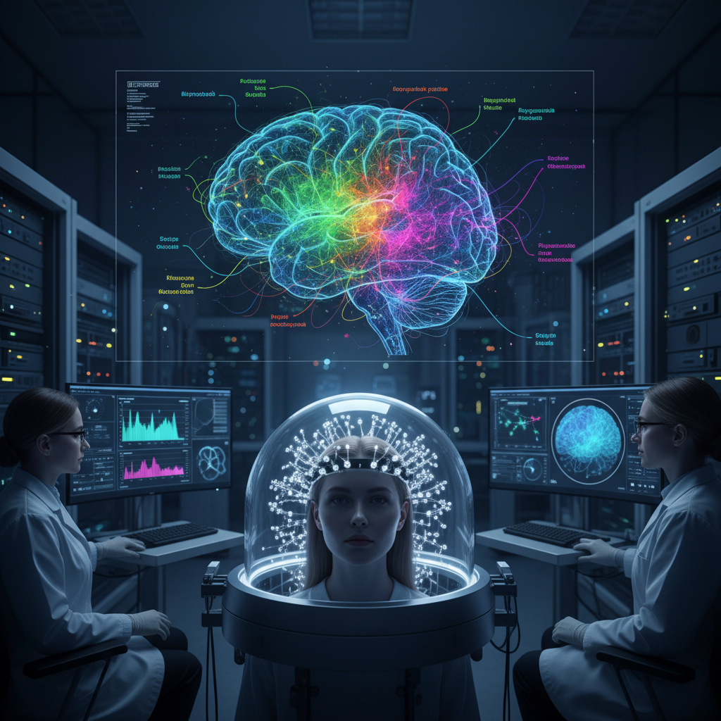
New brain imaging tools are getting better at tracking activity across multiple cell types at once
New brain imaging tools are getting better at tracking activity across multiple cell types at once
One of the oldest technical problems in neuroscience has been surprisingly simple to describe: the closer you look at the brain, the less of it you can see at one time.
For years, researchers have had to choose between high detail and broad coverage. They could zoom in on individual cells or small local circuits with impressive precision, or step back and watch activity unfold across much larger brain regions. Doing both at once has been far harder.
That trade-off matters because the brain does not work at just one scale. Behaviour, perception, attention, movement and internal state all emerge from interactions between small cellular networks and larger brain-wide systems. If scientists can only see one layer at a time, they miss part of the story.
The strongest safe interpretation of the supplied evidence is that newer imaging platforms are improving scientists’ ability to monitor coordinated brain activity across large regions and across multiple cellular populations at the same time. That could help researchers better understand how circuits and behaviours emerge from the interaction of different brain cell types. What the supplied studies do not directly establish, however, is the specific claim that activity was captured across exactly nine cell types at once. The broader advance is well supported; the exact numerical headline is less directly verified by the references here.
Why this kind of imaging matters
The brain is not just a mass of identical neurons firing in unison. It is a layered, highly diverse system made up of many cell populations with different roles. Some cells excite activity, some suppress it, some help regulate timing and coordination, and others support the environment in which signalling happens.
That means understanding the brain increasingly depends on understanding interaction, not just isolated activity. Scientists want to know not only which cells are active, but how local groups of cells connect to larger waves of activity that spread across broader networks.
This is where modern brain imaging has become especially important. The field is moving towards tools that can capture neural dynamics across wider areas while still preserving meaningful cellular detail.
The rise of mesoscale imaging
A key part of this story is mesoscale calcium imaging, which features prominently in the supplied literature.
This approach allows researchers to monitor coordinated neural activity over large fields of view. Instead of observing only a tiny patch of brain tissue, scientists can track patterns spanning broad cortical regions. That makes it possible to study how different areas of the brain activate together, shift with behavioural state, or reorganise during movement, attention or sensory processing.
The supplied evidence supports mesoscale imaging as an increasingly valuable tool for studying widespread circuit dynamics. In practical terms, it gives researchers a more complete picture of how the brain behaves as an integrated system rather than a collection of disconnected parts.
That is a meaningful advance on its own. Large-scale brain activity often contains information that is invisible when researchers focus only on a small local circuit.
Connecting the small scale to the large scale
One of the more compelling findings in the supplied studies is that one research group combined mesoscopic imaging with cellular-resolution two-photon microscopy.
That matters because it links two levels of brain observation that have often been kept separate. On one side is the wide-angle view of cortex-wide dynamics. On the other is the fine-grained view of what individual neurons or small local populations are doing.
According to the supplied evidence, this combined approach showed that both local neuronal correlations and large-scale network activity encode behaviour. That is a powerful result. It suggests behaviour is not represented only in tiny local cell clusters, nor only in broad network states, but across both levels at once.
This pushes neuroscience towards a more integrated model of brain function. To understand behaviour, it may not be enough to study either the micro-scale or the macro-scale alone. The crucial insight may lie in how they interact.
Multimodal recording is deepening the picture
Another supplied study points in a similar direction by showing that simultaneous optical and electrophysiological recording across wide cortical regions can reveal brain dynamics that depend on behavioural state.
This kind of multimodal recording matters because each method sees a different aspect of brain function. Optical imaging can map broad spatial patterns of activity. Electrophysiology adds a more direct view of electrical signalling and timing.
When these methods are used together, researchers can build a richer picture of how the brain is functioning in real time. They are not just seeing where activity is happening, but also how it unfolds, how it relates to changing states, and how local signals fit into distributed network dynamics.
That is especially important for studying behaviour, because behaviour is rarely the product of one brain area acting alone. It reflects coordination across multiple systems that shift from moment to moment.
What the headline gets right
The main strength of the headline is its direction.
The supplied studies clearly support the idea that modern imaging tools are improving scientists’ ability to follow coordinated brain activity across diverse cell populations and spatial scales. They also support the broader claim that this could sharpen our understanding of how neural circuits give rise to behaviour.
In other words, the big story is real: neurotechnology is moving towards a more complete way of seeing the brain. Researchers are increasingly able to connect cellular detail with brain-wide dynamics instead of treating them as separate problems.
That is a major methodological shift, and it could influence how future studies approach everything from sensation and movement to learning, memory and disease models.
Where caution is needed
The caution point is the specific claim about nine cell types at once.
The supplied PubMed evidence does not directly verify that exact statement. Most of the literature provided is focused on mesoscale calcium imaging, cortex-wide dynamics, multimodal recording and the integration of wide-field and cellular-resolution methods. That supports the broader story of multicellular and multiscale imaging progress, but not the precise quantitative claim in the headline.
So the most accurate editorial framing is not that the supplied studies prove a definitive “nine-cell-type” milestone. It is that they support a clear technological direction: brain imaging is becoming more capable of capturing coordinated activity across multiple cellular populations and large brain regions at the same time.
Still largely a preclinical research tool
Another limitation is that these studies are mostly methodological and preclinical, largely in mouse models.
That does not diminish their scientific value. Basic neuroscience often advances through exactly this kind of tool-building. But it does mean the immediate clinical implications are limited.
These imaging systems are not yet telling us how to diagnose neurological disease in routine care, nor do they fully capture how the human brain functions in natural settings. Their current role is more foundational: helping researchers build better maps of how activity is organised across cells, circuits and large-scale networks.
Even then, no method captures everything equally well. Advanced imaging platforms still involve trade-offs among:
- spatial coverage;
- cell-type specificity;
- imaging depth;
- temporal resolution;
- and fine cellular detail.
So while the tools are getting better, they are not all-seeing. Any claim that they fully capture all relevant brain-cell interactions would go too far.
Why this could shape the future of neuroscience
Even with those caveats, the implications for research are significant.
If scientists can better observe how different cell populations participate in distributed brain activity, they may be able to ask more realistic questions about how the brain actually works. Instead of treating behaviour as the output of one region or one cell class, they can study it as the product of interactions across multiple scales.
That could matter for understanding:
- how brain regions coordinate during complex behaviour;
- how local activity patterns influence broader network states;
- how distinct cell populations contribute to perception, action and internal state;
- and how disease-related disruptions spread from circuits to networks.
This does not mean neuroscience is suddenly close to fully decoding the brain. But it does mean the field is developing better tools for studying the brain as a dynamic, interconnected system.
The balanced takeaway
The most responsible reading of the supplied evidence is that new brain imaging approaches are improving researchers’ ability to monitor activity across broad brain regions and multiple cellular populations at the same time, which could deepen understanding of how circuits and behaviours emerge.
Mesoscale calcium imaging supports large-field monitoring of coordinated activity. Combined mesoscopic and cellular-resolution imaging suggests that both local neuronal correlations and large-scale network patterns encode behaviour. Simultaneous optical and electrophysiological recording adds another layer by showing how widespread cortical dynamics shift with behavioural state.
Together, these findings strongly support the broader technological advance behind the headline.
But precision matters. The supplied studies do not directly confirm the exact claim that activity was captured across nine cell types at once, and their immediate relevance remains largely limited to preclinical neuroscience research.
Still, the direction is clear. Brain imaging is becoming less about choosing between a close-up and a wide shot, and more about combining both. That may turn out to be one of the most important shifts in how scientists study the brain: not as isolated cells or isolated regions, but as a living network of many cell types working together.