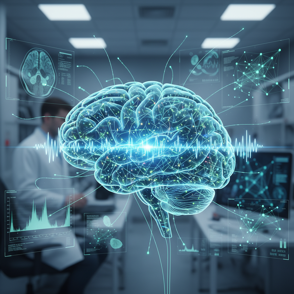
Brain imaging may help explain how deep brain stimulation works in Parkinson’s disease — but the exact mechanism is not confirmed by the evidence provided
Brain imaging may help explain how deep brain stimulation works in Parkinson’s disease — but the exact mechanism is not confirmed by the evidence provided
Deep brain stimulation is no longer a novelty in Parkinson’s disease care. In carefully selected patients — particularly those with persistent tremor, major motor fluctuations or troublesome medication-related dyskinesias — it can reduce symptoms and improve quality of life. What remains much less settled is another question: exactly how does it produce those effects in the brain?
That gap is what makes any headline about imaging “showing” how deep brain stimulation works so appealing. The idea makes sense. Modern neurology increasingly treats Parkinson’s disease less as a problem of one isolated structure and more as a disorder of brain networks. Within that framework, using neuroimaging and connectomics to map circuits and understand neuromodulation is a natural next step.
But there is an important caution. Based on the scientific material provided here, the specific imaging finding in the headline cannot be directly verified. The supplied PubMed papers support the broader plausibility of the story — that imaging and network neuroscience may help explain how deep brain stimulation works in Parkinson’s disease. What they do not do is report the specific study behind the headline or directly establish the exact mechanism being suggested.
What is already known with confidence about deep brain stimulation
The firmest part of this story is not the fine-grained mechanism, but the established clinical role of deep brain stimulation itself.
The treatment involves implanting electrodes in specific deep brain targets and connecting them to a programmable pulse generator. In clinical practice, it is used mainly when motor symptoms no longer respond smoothly to medication, or when medication begins to produce fluctuations and dyskinesias that become difficult to manage.
That matters because it shows deep brain stimulation is not an abstract experimental concept. It is already a real therapeutic tool. The challenge is that its clinical usefulness was established before neuroscience fully worked out every detail of how it reorganises brain activity.
That is not unusual in medicine. Sometimes a therapy proves useful before its mechanism is completely understood. Deep brain stimulation partly fits that pattern.
The shift in perspective: Parkinson’s as a circuit disorder
One of the most important shifts in modern neurology is the idea that conditions such as Parkinson’s disease can be understood in terms of brain circuits, not just local dysfunction.
Within this framework, symptoms such as rigidity, bradykinesia and tremor emerge from disruption across networks linking the basal ganglia, thalamus, cortex and other regions involved in movement planning and execution. That helps explain why stimulating one deep target may produce effects that ripple across motor function more broadly.
The supplied review on brain connectomics supports that view well. It reinforces the idea that neurological and psychiatric disorders are increasingly being studied as network-level conditions, and that this framework can inform neuromodulation research. That point is central here: it does not prove the headline, but it does make its overall direction biologically coherent.
Why imaging seems so promising in this field
If Parkinson’s is at least partly a network disorder, then it makes sense to study those networks with imaging tools. That is where the enthusiasm around functional MRI, tractography, structural connectivity mapping and other modern imaging approaches comes in.
These methods promise something valuable: moving beyond the idea that deep brain stimulation simply “switches on” or “switches off” one region, and instead viewing it as an intervention that alters communication across disease-relevant circuits.
That shift matters because it could help answer both scientific and practical questions at once. For example:
- which networks are linked to the best clinical response;
- why some patients improve more than others;
- how different surgical targets affect different circuits;
- and how unwanted cognitive, emotional or behavioural effects might arise.
That is why the headline feels plausible. Imaging really could help clarify the network logic behind deep brain stimulation. The problem is that, without the key study itself, plausibility cannot be mistaken for direct proof of the specific mechanism described.
The mismatch between the headline and the supplied studies
This is the main editorial caution. None of the PubMed articles supplied directly reports the specific imaging study cited in the headline or clearly explains how deep brain stimulation “acts” in Parkinson’s disease through a newly demonstrated imaging mechanism.
One paper is a general review of Parkinson’s disease. Another offers a broad overview of brain connectomics. A third focuses largely on eating behaviour and neuromodulation, making it only indirectly relevant. Together, they provide useful scientific background. But they do not provide the central evidence the headline implies.
That matters because, without the key supporting study, it is not possible to know:
- which imaging modality was used;
- how many patients were included;
- what networks or regions were identified;
- whether the findings were observational or comparative;
- and how strong the connection was between the imaging results and clinical benefit.
Without those details, any more specific statement about mechanism risks going beyond the available evidence.
What can be said more safely
Even with that limitation, there is still a solid and useful interpretation available. Deep brain stimulation in Parkinson’s disease probably should not be understood as a purely local correction at one anatomical point. The modern framework suggests that it reshapes activity across networks related to motor control, and perhaps beyond.
In that sense, imaging and connectomics are promising because they may help build a more refined map of those networks. They could bring clinical practice closer to a mechanistic explanation that is less dependent on symptom observation alone and more anchored in systems neuroscience.
That reading fits the supplied articles well. What it does not do — and should not do — is pretend that we already know exactly which circuit was modulated, what signal was seen on imaging or how that specific finding explains deep brain stimulation in all patients.
Why this matters even without a final answer
It may sound disappointing to say the exact mechanism is still unresolved. But that does not make the story less important. If anything, it makes it more relevant.
In neuromodulation, understanding mechanism better could have practical consequences. It could help refine surgical targeting, improve stimulation programming, predict response more accurately and perhaps reduce unwanted effects on cognition, mood or behaviour.
So the value of this research agenda does not depend on already having a final answer. It lies in the fact that neurology is moving towards a stage where brain interventions are increasingly understood as the modulation of functional networks rather than simply the stimulation of one spot.
That is an important conceptual change.
The risk of promising more than imaging can yet deliver
It is also worth recognising a common risk in stories like this: treating brain imaging as though it were a transparent window into disease mechanism. In reality, imaging studies often show correlations, connectivity patterns and changes associated with clinical states or interventions. That is valuable, but it is not always the same thing as full causal proof.
Even when a study shows that a certain network changes after stimulation, the next question remains difficult: is that change the direct cause of improvement, an indirect consequence, a related marker or just one part of a larger system? In clinical neuroscience, that distinction is rarely straightforward.
So the safest reading of the headline is as a strong and biologically plausible circuit hypothesis — not as a settled mechanism.
The most balanced reading
The supplied evidence supports the broad idea that neuroimaging, connectomics and network-based neuroscience are increasingly important in understanding Parkinson’s disease and the effects of deep brain stimulation. It also supports the view of Parkinson’s as a circuit disorder, which makes it biologically plausible that imaging studies may help explain how neuromodulation produces clinical benefit.
But the match between the supplied studies and the specific headline claim is weak. None of the papers directly reports the core imaging study or proves the exact mechanism described in the title. That means it is not possible, based on the material here, to say with confidence exactly how deep brain stimulation acts according to that new imaging finding.
The most responsible conclusion, then, is this: modern imaging will probably play an increasingly important role in explaining how deep brain stimulation modulates brain networks in Parkinson’s disease. But with the evidence available here, the headline’s mechanism should still be treated as an emerging and biologically plausible circuit hypothesis, not as an established fact.