A new generation of tools is revealing how neurons and support cells communicate in the brain — but this is still a research advance, not a routine clinical test
A new generation of tools is revealing how neurons and support cells communicate in the brain — but this is still a research advance, not a routine clinical test
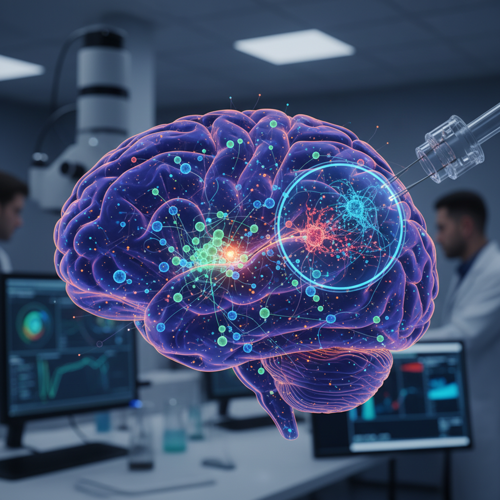
The brain has never worked as a collection of isolated cells. Neurons, astrocytes, microglia, oligodendrocytes, and other cell populations are in constant dialogue, exchanging chemical signals, adjusting metabolism, shaping inflammation, and sustaining circuit function. The problem, for a long time, was technical: science could study brain regions, broad cell classes, or individual pathways reasonably well, but it was much harder to see how different cell types work together at the same time.
That is exactly what this new headline is trying to capture. And in this case, the broad direction is well supported by the evidence provided. Modern single-cell and multiomic methods are making it possible to map how different brain cell types communicate and how those networks shift in ageing and neurological disease.
Still, the wording needs care. The safest version is not that there is now a simple tool that literally “sees” cellular cooperation inside living brains in the way a clinical scan might. What the studies support is something more precise: advanced research platforms can infer and map interaction networks among different brain cell types with unprecedented resolution, generating valuable clues about brain function, ageing, and disease.
What has changed in brain research
For decades, neuroscience advanced through methods that were enormously useful but limited in resolution for questions like this. Many studies measured average activity across whole tissue samples, entire brain regions, or broad cell populations. That taught researchers a great deal about anatomy, electrical activity, and disease, but it also blurred some of the finer cellular conversations taking place underneath.
Single-cell and multiomic methods have changed that landscape because they allow researchers to separate individual cells and examine, in much greater detail, which genes are active, which regulatory elements may be controlling them, and what kinds of signals different cells may be sending to one another.
In practical terms, that means moving from a relatively blurred portrait of the brain to something closer to an interactive map, where scientists can ask:
- which cell types are present;
- what each one is expressing;
- which regulatory pathways appear to shape those states;
- and which cell pairs may have the potential to communicate through specific molecules.
What the strongest evidence shows
One of the key references provided describes a large single-cell genomics resource in the human brain. That work identified cell-type-specific regulatory elements, as well as expression quantitative trait loci and cell-to-cell communication networks across many brain cell types.
That matters because it connects several layers of biology at once. It is no longer just about which genes are turned on in a given cell. It is about beginning to understand how genetic variation, gene regulation, and cross-talk between cell types come together.
That is what makes the headline broadly plausible. The brain is not simply a sum of separate cells; it is a system in which the state of one cell population may influence many others.
The value becomes clearer when disease is involved
Another of the supplied studies, based on Parkinson’s disease brain tissue, showed something even more concrete: altered neuron-astrocyte interactions together with enhanced neuroinflammation.
That is important because it moves beyond a purely descriptive map. It shows that these tools can detect changes in how different cell types relate to one another in a real human disease setting.
That helps shift neuroscience beyond an overly neuron-centred view. Instead of treating disease only as neuronal death or dysfunction, these methods suggest that part of the problem may lie in the relationships between neurons and support cells, especially in inflammatory and metabolic processes.
In other words, disease may not belong to just one cell type. It may reflect a disrupted cellular network.
Ageing also appears to be a communication problem
The third reference reinforces the same idea in another context: brain ageing in mice. That work mapped ligand-receptor interactions across many cell populations, suggesting that ageing is not just a matter of losing cells or reducing global function. It also involves changing the way different cell types signal to one another.
That matters because it brings ageing and disease closer together. Often the line between the two is not drawn by completely different mechanisms, but by different intensities and patterns of cellular dysregulation.
If brain ageing involves coordinated but cell-specific changes in communication, then tools capable of mapping those changes could become very important for understanding why some brains age with greater resilience while others move earlier towards neurodegeneration.
What these tools actually “see”
The headline says a new tool can see how different brain cell types “work together”. That is partly true, but it needs translating carefully.
These methods do not literally watch cells cooperating in real time inside a living brain in the way a camera would capture behaviour. What they do is generate maps of gene expression, molecular regulation, and ligand-receptor pairing that suggest communication potential and coordinated states across cell populations.
That is extremely powerful for research, but it remains a layer of inference. To prove exactly how those cells behave functionally, in which direction signals move, and what biological effects follow, additional experiments are usually still needed.
So “see” should be understood here in a scientific sense: to map, infer, and reconstruct probable cellular relationships at high resolution — not to directly observe full cooperation in a literal, clinically actionable way.
Why this could matter for brain disease research
Even with those cautions, the potential is substantial. Neurological and psychiatric diseases often involve multiple cell types at once. Inflammation, metabolism, synaptic plasticity, oxidative stress, tissue repair, and vascular regulation rarely belong to one cell population alone.
By mapping communication networks, these tools may help answer questions such as:
- which cells initiate pathological responses;
- which respond secondarily;
- which molecular signals become too strong or too weak;
- and which interactions may eventually become more precise therapeutic targets.
That does not mean treatments are suddenly close at hand. It does mean the logic of the research is becoming more refined.
What this story gets right
The story gets something important right by highlighting that new tools are making it possible to map how different brain cell types interact. That is one of the most important shifts in contemporary neuroscience.
It is also right to suggest that this kind of advance may sharpen understanding of brain ageing and disease. The supplied evidence supports that direction consistently, across human brain tissue and animal models, and across both neurodegeneration and ageing biology.
It is also right to treat cell-to-cell communication as a central part of brain function rather than a secondary detail.
What should not be overstated
At the same time, it would be an overstatement to describe this as one simple stand-alone tool already ready for widespread hospital or clinic use. The evidence describes advanced research platforms, not routine clinical technologies.
It would also go too far to say that these methods prove exactly how cells behave over time in living human brains. Some of the key findings come from postmortem human tissue, mouse models, or inferred communication from molecular profiles.
That limits direct causal interpretation. The resulting maps are rich and informative, but they are not the same thing as a complete demonstration of dynamic cell behaviour in real time.
What this may mean for the future
The most promising impact may lie in integration. As these techniques are combined with imaging, physiology, human genetics, and functional experiments, neuroscience may move from a more descriptive phase to one in which the brain’s multicellular organisation is understood with much greater precision.
If that happens, the payoff could be large: sharper biologic classification of disease, more realistic disease models, better target selection, and a less simplistic view of the brain as an organ dominated only by neurons.
Even so, that future depends on validation, reproducibility, and careful translation. The path from research platform to clinical application is usually a long one.
The most balanced reading
The safest interpretation is this: new single-cell and multiomic methods are making it possible to map, in far greater detail, how neurons and glial cells communicate, offering valuable clues about brain ageing and neurological disease.
The supplied evidence supports that direction well. A major human brain single-cell genomics resource identified communication networks across many cell types; Parkinson’s disease brain analyses revealed altered neuron-astrocyte interactions together with increased neuroinflammation; and ageing mouse brain studies mapped changing ligand-receptor relationships across multiple cell populations.
But the limits remain decisive: these are sophisticated research platforms, not routine tests; inferred cell-cell communication still needs functional validation; and the data do not mean that cellular cooperation is being directly, literally, and clinically observed in living brains.
In short, the headline points to a real and important scientific advance. The most solid message is not that neuroscience has suddenly found a magic tool that can fully watch the brain at work, but that it now has much more powerful ways to reconstruct how different cell types jointly shape ageing and disease.